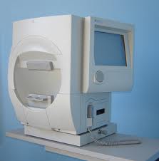
Visual Fields Testing
 A visual field test is a method of measuring an individual’s central and peripheral (side) vision. Visual field testing is most frequently used to detect any signs of glaucoma damage to the optic nerve. In addition, visual field tests are useful for detection of central or peripheral retinal disease, eyelid conditions such as ptosis or drooping of the eyelid, optic nerve disease, and diseases affecting thePerimeter.
visual pathways within the brain. Many eye and brain disorders can cause peripheral vision problems and visual field abnormalities. Brain abnormalities such as those caused by strokes or tumors can affect the visual field. In fact, the location of the stroke or tumor in the brain can frequently be determined by the size, shape and site of the visual field defect.
To do the test, you sit and look inside a bowl-shaped instrument called a perimeter. While you stare at the center of the bowl, lights flash. You press a button each time you see a flash. A computer records the spot and at the end of the test, a printout shows if there are areas of your vision where you did not see the flashes of light. These are areas of vision loss. If you are diagnosed with a particular disorder or disease such as glaucoma, visual field testing will become a routine part of your treatment. After a baseline has been established, visual field testing is repeated every 6 to 12 months to monitor for change. Visual field tests plays a critical role in helping the doctor follow your condition.
