Advanced Diagnostic Testing
Vision Health Institute is equipped with the most advanced diagnostic testing equipment to ensure our patients that they are receiving the most thorough and complete eye exam possible. Our highly-trained staff are ready to answer any questions that you might have. The following diagnostics are just some of the tests you will receive during your eye exam. Schedule your exam today!
Pachymetry
This instrument uses ultrasound to measure corneal thickness. Measurements of the cornea are significant in determining if you are a candidate for refractive laser surgery. Additionally, thin corneas can lead to falsely low pressure readings and thick corneas can lead to falsely high pressure readings. This test is done just once to create a baseline for future testing. Pachymetry may be needed if you are being considered for refractive corneal surgery or possibly glaucoma.
Humphrey Visual Field Testing (peripheral vision or perimetry)
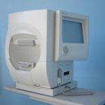 Your visual field is the area you are able to see in front of you without moving your eyes, including your side vision. There are several different methods of testing the visual field. Using a few different methods of testing, your vision is “mapped” to determine what you see at the edges (periphery) of your visual field. This helps to diagnose several conditions. The patient sits in front of a concave dome with a target in the center. The eye that is not being tested is covered. A button is provided to the patient to be used during the exam. The patient is asked to focus on the target at the center. The computer shines lights (of varying intensity) on the inside dome and the patient clicks the button when a light is seen. The computer automatically maps and calculates the visual field. Read more about visual field testing…
Amsler Grid Testing
Amsler grid testing is often recommended for patients with macular diseases. The Amsler grid is an effective way for patients to monitor for changes in their central vision. Patients with significant macular disease are often advised to check the grid daily. The Amsler grid is simply a small square of graph paper with a dot in the center. Patients often apply the grid to a mirror, refrigerator, or some other prominent place in the house where they will see it and remember to check it daily.
Digital Slit Lamp Imaging
A high- resolution digital camera attached to the Slit Lamp Microscope allows the doctor to have a permanent photo-image of the eye condition. This allows the doctor to manage external diseases and conditions of the eye. Digital images are used to compare progressive conditions of the eye to better determine treatment options. Conditions may include eyelid diseases, corneal ulcers, degenerations, and dystrophies.
Retinal Scanning Laser (OCT)
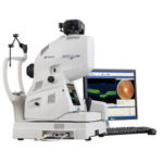 Optical Coherence Tomography (OCT) is a non-invasive imaging technology used to obtain high resolution cross-sectional images of the retina. This allows the doctor to see and measure very slight changes in the retina and optic nerve. The OCT uses light waves in a process similar to ultrasound to test for a number of retinal conditions, which could include glaucoma, Age-Related Macular Degeneration (AMD), post-cataract surgery edema, or other retinal changes. The OCT provides an extremely detailed understanding of retinal eye conditions and allows monitoring of treatment. Read more about OCT…
Retinal Photography
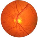 This non-invasive High Definition Photograph generates an instantaneous, digital image of the retina, revealing important information for the comprehensive evaluation of systemic and ocular health. It provides a permanent digital documentation, which allows accuracy in diagnosing disease of the retina and optic nerve. Provides the ability to measure progression of glaucoma and track the effectiveness of medications. Read more about digital retinal photography…
Genetic Testing For Age-Related Macular Degeneration
We now have the capability to offer to our patients the ability to Genetically test them for Macular degeneration. The test results will predict the probability of the patient to develop AMD (Age-related Macular Degeneration).